














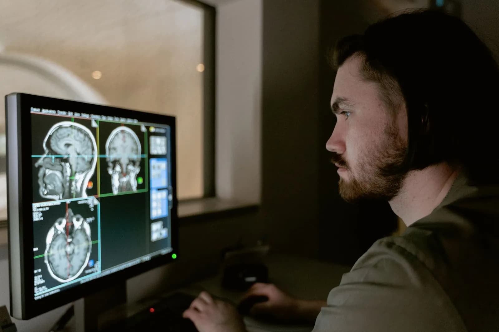
Medical image analysis software doesn't diagnose anyone. It looks at a scan, marks what it thinks is abnormal, and hands that mark to a radiologist who decides whether it matters. Understanding that division of labour is the fastest way to judge any product in this category.
On September 18, 2026, Diagnostic Imaging reported that DeepHealth's Chest XRay software had received FDA 510(k) clearance for adjunctive chest X-ray assessment. It aids detection and localisation of lung nodules, mediastinal abnormalities, consolidation and pneumothorax, and DeepHealth describes it as one of the first tools of its kind built on a foundation model.
What the software does with a scan
It reads the image data, looks for patterns it was trained to associate with a finding, and flags the location. The DeepHealth clearance is a good example: the software points to a suspected nodule or a collapsed lung on a chest film, and the radiologist reviews the flag alongside the image.
Note the word adjunctive. Cleared medical image analysis software of this kind sits beside the reader rather than replacing the reading. Everything downstream, from how it's built to how it's tested, follows from that.
The software also has to cope with a scan arriving from a machine it has never met. That's where a shared file standard earns its keep.
Where DICOM and your PACS fit
Medical image analysis software only works on the images it can receive, and DICOM is how they arrive. The DICOM standard is recognised by ISO as ISO 12052 and is built into nearly all radiology, cardiology imaging and radiotherapy equipment, from X-ray and CT to MRI and ultrasound.
First published in 1993, it sets the formats that let images be shared without losing the quality patient care needs. For a buyer, that makes DICOM support the first checkbox, not a differentiator.
The harder question is how a new tool connects to the systems already holding your studies. That means pulling images from your archive, returning results where radiologists already look, and not asking anyone to open a second application. Integration architecture usually decides how smooth a rollout feels long before the model's accuracy does, which is our own view from building connected healthcare products rather than a measured figure.
Does medical image analysis software make readers more accurate?
Not reliably. In a study written up by EMJ in September 2026, researchers at the Technical University of Munich had five radiologists read the same chest X-rays without AI and then with each of four commercial tools. The set covered 1,200 patients, and the findings were published in Academic Radiology.
Reading got faster for three of the five radiologists, and four of the five reported more confidence. Overall diagnostic accuracy didn't improve.
For pleural effusions and pulmonary nodules, some reader and tool pairings did worse than the reader alone, which the researchers attributed to more false positives. They also raised automation bias, the risk that a reader accepts an incorrect AI finding, and recommended testing each tool in the setting where it will be used.
The caveats matter here. Five readers with one to six years of experience is a narrow group, and the study can't tell you how a tool performs for a specialist or in your hospital. But it's enough to retire the assumption that adding AI automatically improves the read.
What speed gains look like in practice
The time saved was small per case. Median reductions of 6 to 17 seconds were reported for the readers who sped up, so the benefit arrives through volume, not through any single study.
A busy reading list is where that adds up. A quiet one may never notice.
That framing changes what you should measure. Track turnaround and the number of follow-up recommendations before and after go-live, not just the vendor's headline sensitivity.
Regulation: what an FDA clearance tells you
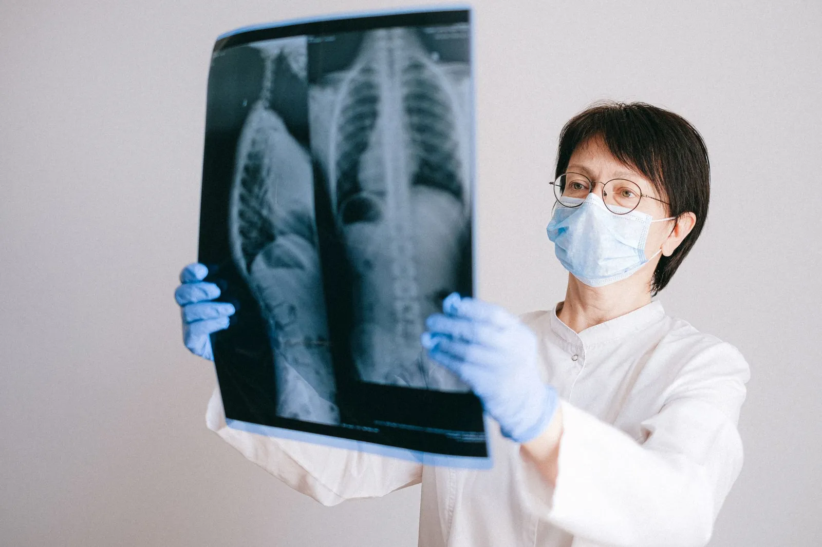
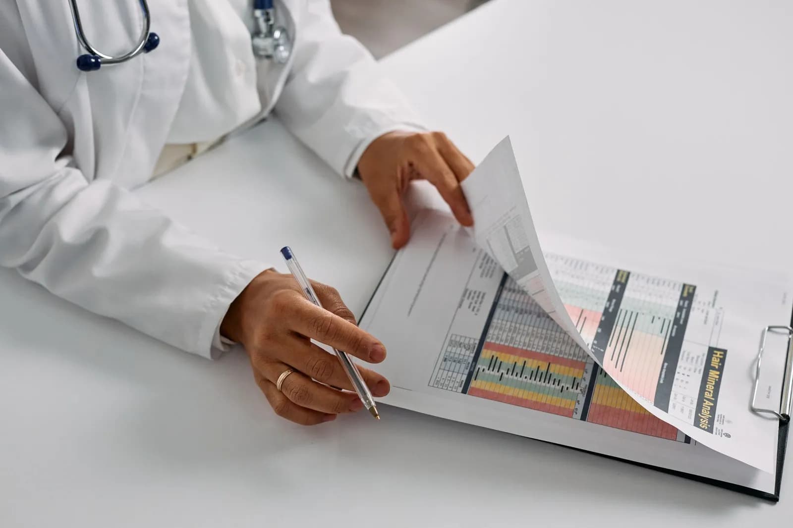
Photo by Anna Shvets on Pexels
It tells you the product met the agency's premarket requirements for a stated use, nothing about whether it will suit your workflow. The FDA's list of AI-enabled medical devices is the place to check a vendor's claim, and in the version we read the most recent final decision was dated 06/29/2026 with radiology the most common panel.
Two limits are worth knowing. The FDA says the list isn't comprehensive, because devices were found mainly through AI-related terms in their authorisation summaries. And the intended use on the clearance is narrow: a chest X-ray tool cleared for adjunctive assessment of certain findings isn't cleared for anything else.
If you are building rather than buying, the clearance route shapes the whole product plan, and it's a conversation to have with regulatory counsel before design starts. Our guide to AI diagnostics software covers the build side in more depth.
Questions to put to a vendor
A demo of any medical image analysis software shows the best case. These questions show the average one:
- Which exact findings and imaging types does the clearance cover?
- How does the tool connect to our PACS, and where does the result appear for the reader?
- What happens to false positives on our own cases, and can we trial it on them before buying?
- Who sees AI marks first, the reader or a worklist, and can readers turn them off?
- Where are images processed and stored, and who has access?
The third question comes straight from the Munich results. A tool that adds false positives for one finding can erase its own speed gain, so ask to see the trade-off on data that resembles yours.
Building your own imaging workflow around AI
Many teams don't want a single vendor's viewer. They want results inside a portal, a clinician app or an existing record system, with the AI as one component. That's a product design and integration job, and it's the kind of connected-care work covered in our piece on healthcare IoT software.
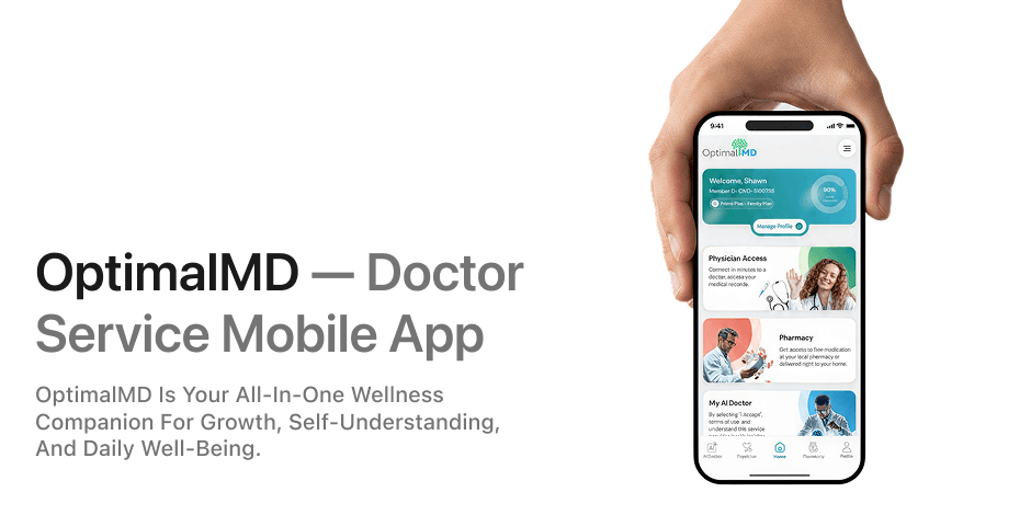
When we built OptimalMD's digital product, we took it from design through development to launch, covering the website, the members portal and the mobile app, as described in the OptimalMD case study. Imaging wasn't part of that project, but the lesson carries: the interface people use every day is what decides whether a clinical tool gets adopted.
If you're scoping something similar, our AI and automation service and apps and SaaS work are the two places to start. For budget planning, see our breakdown of the cost of custom AI solutions.
Start with one narrow use, one imaging type and one reading workflow. Prove it on your own cases, then widen it.
Frequently asked questions
Does medical image analysis software replace radiologists?
No. The FDA-cleared chest X-ray tool in the news this September is cleared as an adjunct, meaning it assists a radiologist's assessment. The radiologist still makes the call.
What file format does the software need?
DICOM, the international standard for medical images and related information, built into nearly all X-ray, CT, MRI and ultrasound equipment. A tool that can't work with DICOM won't fit a normal imaging setup.
Can AI make a reader worse?
In the Munich chest X-ray study, some reader and tool combinations were less accurate for pleural effusions and nodules because of added false positives. That's why testing a tool on your own cases matters before rollout.
How do I check whether a tool is FDA cleared?
Search the FDA's list of AI-enabled medical devices, which links each entry to its clearance record. The agency notes the list isn't comprehensive, so also ask the vendor for the submission number.
Cover photo by MART PRODUCTION on Pexels
Sources
- Foundation model-based chest X-ray AI software gets FDA nod — Diagnostic Imaging
- AI Speeds Chest X-Ray Reading but Not Accuracy — EMJ